ITK Release 4/Data Collection: Difference between revisions
From KitwarePublic
Jump to navigationJump to search
| Line 24: | Line 24: | ||
| Raghu Machiraju || 10 Gb || TIFF || | | Raghu Machiraju || 10 Gb || TIFF || | ||
|- | |- | ||
| Ziv Yaniv || 10Gb || DICOM, meta ( | | Ziv Yaniv || 10Gb || DICOM, meta (e.g. segmentation label maps), bmp/jpg/avi (2D frame grabbed US and x-ray fluoroscopy images)|| | ||
|- | |- | ||
| Brad Lowekamp || 10 Gb || TIFF ? || | | Brad Lowekamp || 10 Gb || TIFF ? || | ||
Revision as of 19:19, 15 December 2010
Overview
Testing Data
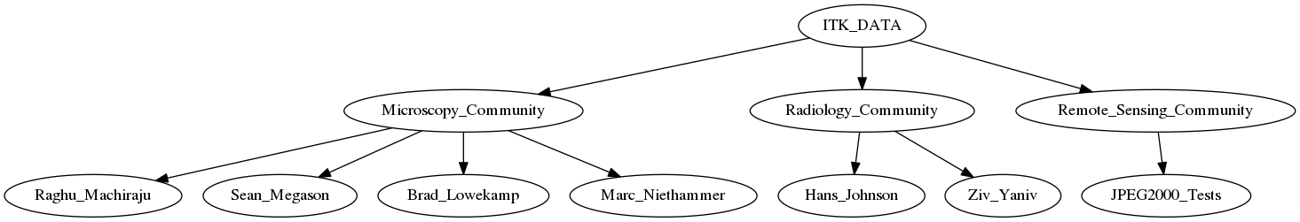
MIDAS Structure
- Communities (recursive)
- Collection
- Items
- Resources
- Bitstreams
- Bitstreams
- Resources
- Items
- Collection
Expected Data
| Group | Expected Amount of Data | File Formats | Features |
|---|---|---|---|
| Raghu Machiraju | 10 Gb | TIFF | |
| Ziv Yaniv | 10Gb | DICOM, meta (e.g. segmentation label maps), bmp/jpg/avi (2D frame grabbed US and x-ray fluoroscopy images) | |
| Brad Lowekamp | 10 Gb | TIFF ? | |
| Hans Johnson | 10Gb | DICOM ? Nifti ? | |
| Sean Megason | 10Gb | TIFF ? | |
| Marcel Pratwsaw | 10Gb | ?? | |
| Marc Niethammer | 10Gb | TIFF |
MetaData
Medical
- physical quantities associated with image acquisition apparatus (e.g. camera calibration for endoscopy and x-ray, US calibration).
- reason for scan (e.g. suspected liver tumor), should be in header but often is not.
- contrast phase (e.g. pre contrast, arterial phase).
- respiratory phase (e.g. end inspiration, end expiration).
- apparatus used to acquire images when not specified in DICOM (e.g. xyz frame grabber).
- rigid transformation data acquired with a tracker (pose of US/endoscope relative to tracking system, required for positioning multiple 2D images in 3D space)
Structure
Suggested Organization
MIDAS Installations
- http://www.insight-journal.org/midas/
- http://www.insight-journal.org/midas/community/view/22 (Miscellaneous collection of ITK related data)
- http://www.insight-journal.org/rire/ (RIRE: Retrospective Registration Evaluation)
- http://midas.kitware.com/
- http://midas.kitware.com/community/view/7 (ITK Testing Data)
- http://midas.kitware.com/community/view/22 (Tutorials: Virtual Appliances > 2Gb)